- -5%
- New

Authors: Daniel Vincent, Paul Winston
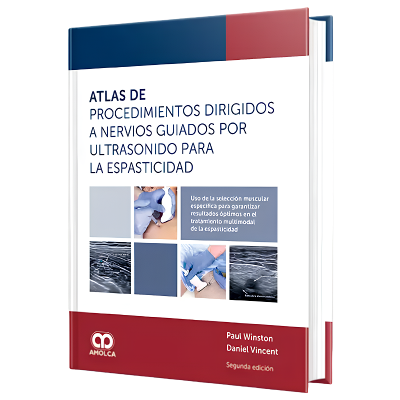
The ATLAS of proceedings directed at ultrasound guided nerves for spasticity, in its second edition, is the optimal work tool for all those who wish to improve clinical outcomes and offer optimal attention to patients with spasticity. Diagnostic nervous blockage is a technique that allows you to efficiently select the most suitable muscles and offer optimal treatment, from botulinum neurotoxin to phenolization, passing through surgery and new interventions.
Shipping From: Spain
Characteristics of the book:
Accurate, clear and clinic-oriented: Chapters include authors and international experts that describe numerous issues, from current definitions of spasticity, an explanation of diagnostic nerve blocks and current concepts in surgery, Chemorenervation and new interventions.
Detailed graphic demonstrations: high quality images illustrate the location of the trunks and branches nervous, as well as muscle action, injection points and the techniques of the injection protocols for each desired muscle or muscle group.
Brief and concise: Clinical practice-oriented techniques are approached and visualized accurately in a compact format. The book is ideal for consulting and optimizing your injection technique.
1. 1.1. About the authors 1.2. Thanks 1.3. How to use this atlas 1.4. Change in the focus of clinical measurement for spasticity
2. Introduction 2.1. Introduction to diagnostic nerve blocks 2.2. History of Blocks Engines and Recommendations in France 2.3. The need for a multidisciplinary collaborative atlas 2.4. Collaborative attention focused on the patient in the neuroortopedia 2.5. The role of anesthesiologist in interdisciplinary care of spasticity 2.6. Spastic Extremities Positions
3. Ultrasound principles 3.1. Orientation of the ultrasound transducer 3.2. Space relationship between nerves, arteries and veins 3.3. The surface reference points of the nervous branches show variation 3.4. The individual anatomy varies. The benchmarks are fundamental 3.5. Surgical neurectomy 3.6. Three-dimensional images of nerves by ultrasound 3.7. Evaluation of muscle changes with ultrasound in spasticity: Use of the modified Heckmat scale 3.8. Tracking a proximal to distal nerve: medium nerve from the round pronator to the intrinsic muscles 3.9. Display of the injected liquid 3.10. Stimulator parameters 3.11. Ergonomic aspects related to nerve blocks guided by ultrasound for spasticity
4. Procedures directed to the nerves 4.1. Chemionourlysis with phenol and ethanol 4.2. CrioneRolysis: a new minimally invasive percutaneous therapy for the treatment of spastive limbs 4.3. The algorithm lives. Use of diagnostic nerve blocks to treat pain again on the shoulder by hemiplejia after a stroke 4.4. Neurectomy for the spasticity of the upper extremity
5. Procedures directed to the nerves of the upper limb 5.1. Shoulder in adduction and internal rotation 5.2. Pectoral muscles and neurovascular structures 5.3. Lateral pectoral nerve: Pectoral muscle 5.4. Medial pectoral nerve: minor pectoral muscle 5.5. ToroCodorsal nerve: wide dorsal muscle 5.6. Espastic elbow in flexion 5.7. Musculocutaneous nerve: brachial biceps muscle 5.8. Musculocutaneous nerve: brachial muscle 5.9. Radial nerve: Braquioradial muscle 5.10. The wrist and fingers: anatomical guide and new use of nervous block with phenol in the distal region of the upper extremity 5.11. Anatomy of the motor branches of the muscles of the higher extremity most involved in the spasticity 5.12. Evaluation of the spastic hand by diagnostic nerve blocks 5.13. Medium nerve: Round pronous muscle 5.14. Medium Nerve: Radial Flexor Muscle Carp 5.15. Medium nerve: Surface flexor muscle of the fingers 5.16. Medium nerve: from the interopeous nerve before the deep finger muscle of the fingers 5.17. Medium nerve: of the interopeous nerve before the long flexor muscle of the thumb 5.18. Medium nerve: from the interherse nerve before the square pronator muscle 5.19. Of the median nerve to the intrinsic muscles: lumbricals 1 and 2, short thumb abductor, short thumb flexor, thumb opponent 5.20. Cubital nerve: Cubital Flexor Muscle of Carpo 5.21. Cubital nerve: deep flex-finger muscle 5.22. Cubital rib: From the deep ulnar branch to the intrinsic muscles - part 1 5.23. Cubital nerve: from the deep cubital branch to the intrinsic muscles - part 2 5.24. Radial nerve: Long and short radial tank muscles of carp 5.25. Radial Nerve: Cubital Extensor Muscle of Carpo 5.26. Radial nerve: Fingering extender muscle
6. Procedures directed to the nerves of the lower extremities 6.1. Sealing nerve 6.2. Nerve Shutter, Previous Division: Branch That Innervates To Muscle ADDUCOR LONG 6.3. Nerve Shutter, Previous Division: Branch that Inerta to the Short Aductor Muscle 6.4. Nerve Shutter, Previous Division: Branch that Inerta to Muscle Gracilis 6.5. Nerve Shutter, Previous Division: Branch that inerty to pectine muscle 6.6. Nerve Shutter, Rear Division: Branch that Ineve to the Main Aductor Muscle 6.7. Femoral nerve 6.8. Femoral nerve: Straight muscle femoral 6.9. Sciatic nerve 6.10. Sciatic nerve: Bicep muscle femoral 6.11. Sciatic nerve: semimembranous muscle 6.12. Sciatic nerve: semi-reinstinous muscle 6.13. The role of diagnostic nerve blocks for the treatment of spastic foot 6.14. Ultrasound of the tibial nerve 6.15. Surgical anatomy of tibial nerve 6.16. Trunk and branches of the tibial nerve 6.17. Tibial nerve: Gastrocnemus muscle 6.18. Tibial nerve: Soleo muscle 6.19. Tibial Nerve: Rear Tibial Muscle 6.20. Tibial nerve: long flexor muscle of the toes 6.21. Of the tibial nerve to the intrinsic muscles: medial and lateral planting nerves

Data sheet
Specific References
Authors: Daniel Vincent, Paul Winston
The ATLAS of proceedings directed at ultrasound guided nerves for spasticity, in its second edition, is the optimal work tool for all those who wish to improve clinical outcomes and offer optimal attention to patients with spasticity. Diagnostic nervous blockage is a technique that allows you to efficiently select the most suitable muscles and offer optimal treatment, from botulinum neurotoxin to phenolization, passing through surgery and new interventions.