- New
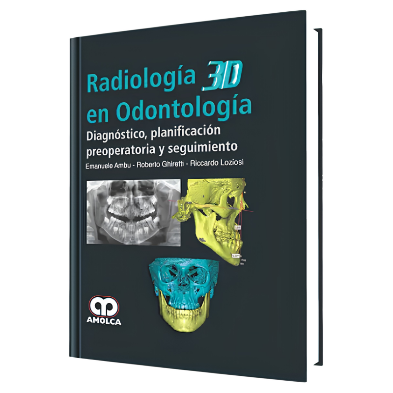

Author: Emanuele Ambu
The conical beam computed tomography (TCHC), is an extraordinary advance for dentistry. With it a new border opens, and allows us to make an accurate diagnosis in those cases where traditional tools were not enough. In daily practice, we are generally facing situations of radiographic exploration and symptoms of our patients who do not coincide, or can even take us to multiple scenarios with different diagnoses. When I look at a traditional X-ray, I always think, in fact, that it is a two-dimensional image of something that has actually has three dimensions. Finally, thanks to the TCHC we have the lacking dimension, which increases our knowledge exponentially.
Shipping From: Spain
When reading the text, we can see the enthusiasm and passion with which the authors elaborated this work. Each chapter is a source of information, every detail was carefully examined, and each clinical case has been presented extensively.
The introductory chapters provide the knowledge and basic tools for the professional to understand the TCHC.
The rest of the book is an extremely pleasant atlas that includes the use of the TCHC in clinical and surgical dentistry, and describes in detail not only the diagnostic phase, but also the operational use to plan the individual cases and control the future outcome.
The work is scheduled to be consulted many times, every day, it is very useful for those who approach this new dimension of dentistry and require a guide. On the other hand, the authors explain, in a very simple way, the concepts that are not at all simple, demonstrating their experience and deep knowledge of matter.
Chapter 1.- Discovery of x-rays to the advent of digital tomography.
Chapter 2.- Principles of 3D Radiology
- Traditional radiological technique and its digital form.
- Three-dimensional radiology: basic theoretical principles.
- Structure and characteristics of 3D radiological systems.
- Acquisition - Reconstruction - Visualization.
Chapter 3.- How to choose a suitable system to the needs of the dentist.
- Clinical requirements, Risk of radiation, definition of the image.
- Criteria to choose an "ideal system" and FOV for any clinical practice.
- Choice of a system based on our daily practice.
- The Alara principle and choice of a system based on the dose of radiation to the patient.
- Legal aspects - Conclusion.
Chapter 4.- Radiological anatomy of the oral cavity and adjacent areas.
- Axial plane - Sagittal plane - coronal plane.
- Examination of the upper respiratory path.
Chapter 5.- Three-dimensional rendering of the models using the TCHC data.
- of virtual models to real models.
- Clinical use of models processed by 3D rendering.
- Use of 3D rendering for communication with patients.
Chapter 6.- Use of the TCHC in dentistry.
- Introduction - Implants - Endodontics.
- Dental Traumatology - Oral Surgery.
- Periodontics - Orthodontics.
- Sinus pathologies of odontogenic origin.
- Bibliographic references.
Alphabetical index

Data sheet
Specific References
Author: Emanuele Ambu
The conical beam computed tomography (TCHC), is an extraordinary advance for dentistry. With it a new border opens, and allows us to make an accurate diagnosis in those cases where traditional tools were not enough. In daily practice, we are generally facing situations of radiographic exploration and symptoms of our patients who do not coincide, or can even take us to multiple scenarios with different diagnoses. When I look at a traditional X-ray, I always think, in fact, that it is a two-dimensional image of something that has actually has three dimensions. Finally, thanks to the TCHC we have the lacking dimension, which increases our knowledge exponentially.